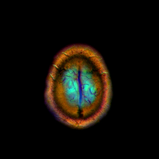
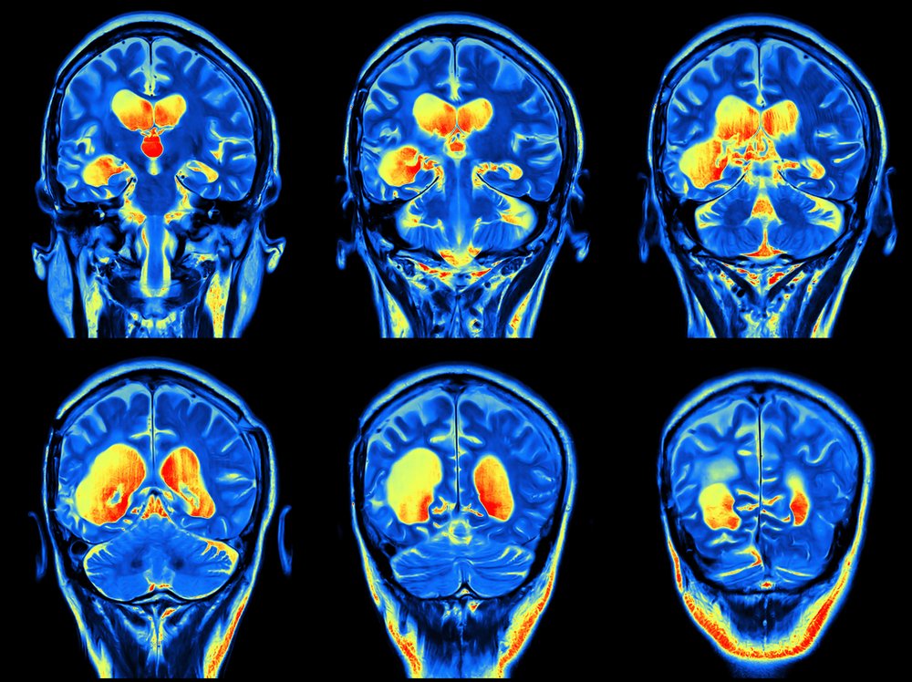
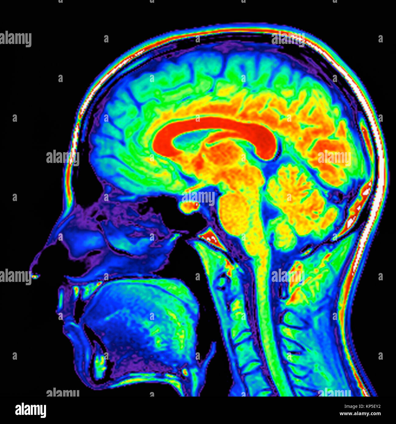
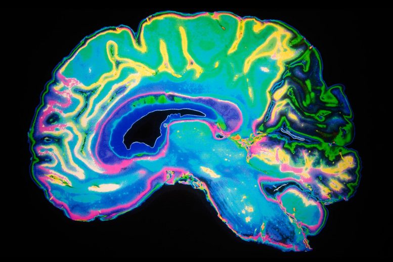
Nevit's blog Color MRI of the Brain
Magnetic resonance imaging (MRI) is a medical imaging technique that uses a magnetic field and computer-generated radio waves to create detailed images of the organs and tissues in your body. Most MRI machines are large, tube-shaped magnets. When you lie inside an MRI machine, the magnetic field inside works with radio waves and hydrogen atoms.
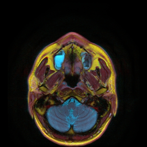
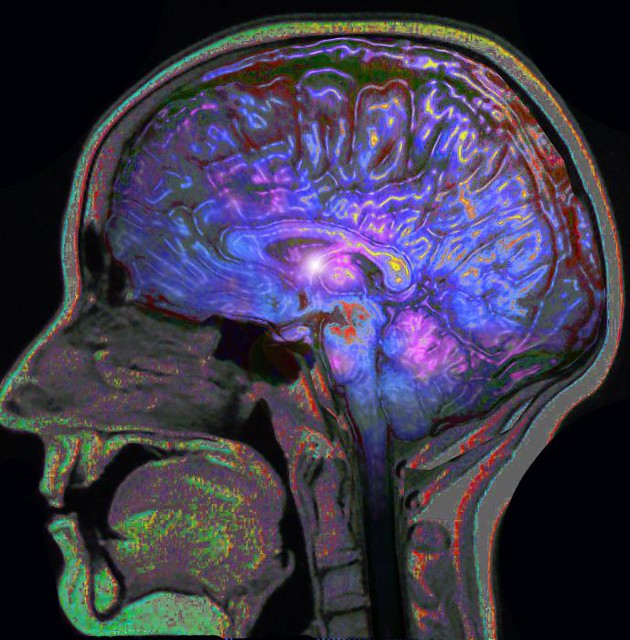
Nevit's blog Color MRI of the Neck
Browse 14,600+ color mri stock photos and images available, or start a new search to explore more stock photos and images. Sort by: Most popular human brain Brain activity,Human brain damage,Neural network,Artificial intelligence and idea concept MRI scan of brain Brain MRI scan. Scanning of brain's magnetic resonance image..
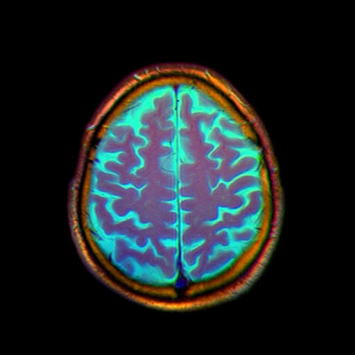
Nevit's blog Color MRI of the Brain
Why do we need to use MRIs? Generally, MRI is used less commonly than plain films and CT scans. They are often reserved for superior viewing of soft tissues. MRI is particularly helpful in patients with suspected neurological or musculoskeletal pathology, however, they can be used in many other specialities too.

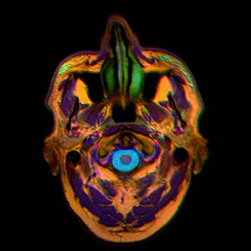
Nevit's blog Color MRI improvements
Diagnostic Imaging Central Scheduling Phone: 603-663-2180 What do MRI Images look like? Are they in color? Here are some examples below of images taken at our facilities. The majority of our scans are done in gray scale however some of our specialty exams offer color imaging as a way to aid the Radiologist using special computers.

Nevit's blog Color MRI of the Brain
Adding "Color" to MRI MRI may be widely used, but the technology is still essentially where black and white film was in the early 20th century. Researchers have now figured out a way to add the equivalent of color to MRI. The advance could help doctors tell different structures and types of cells apart in images of your insides.
Nevit's blog Color MRI of the Brain
The two basic types of MRI images are T1-weighted and T2-weighted images, often referred to as T1 and T2 images. The timing of radiofrequency pulse sequences used to make T1 images results in images which highlight fat tissue within the body.

Nevit's blog Creating Color MRI images with Osirix Color MRI Plugin
Now, a U.S. research team is developing injectable micromagnets which could bring color to MRI pictures. The researchers also point out that these 'new micromagnets also could act as smart.
ms symptoms but normal mri Captions Definition
Synthetic images resembling brain dynamic-contrast enhanced MRI consisting of scaled mixtures of white, lumpy, and clustered backgrounds were used to assess the performance of a rainbow ("jet"), a heated black-body ("hot"), and a gray ("gray") color scale with display devices of different quality on the detection of small changes in color intens.

Know Your Neuro, Save A Life Ophthalmology Advisor
A color MRI image holds much more information than grayscale image; so a proper segmentation technique is essential to recognize the affected portion accurately. MRI image segmentation is mainly used to identify tumors, classification of tissues and blood cells, multimodal registration, etc.
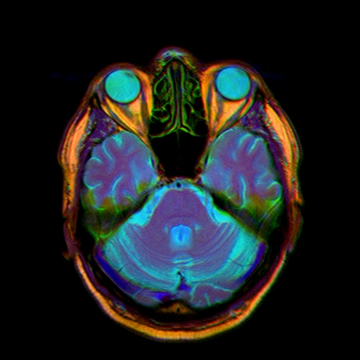
Nevit's blog Color MRI of the Brain
Browse 12,200+ color mri pictures stock photos and images available, or start a new search to explore more stock photos and images. Sort by: Most popular human brain Brain activity,Human brain damage,Neural network,Artificial intelligence and idea concept Medical MRI Scan Medical MRI Scan on digital screen MRI scan of brain
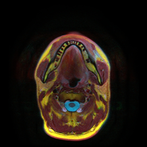
Nevit's blog Color MRI of the Neck
The colorization network was trained on 1707 Visible Human cryo-anatomical images and 50,094 MRI images, while the segmentation network was trained on 94,000 Oasis MRI images with their labels. Additionally, to prevent the image background from interfering with the target colorization object in the training process, we remove the background.

Elliot Hospital MRI What do MRI Images look like?
Citation, DOI, disclosures and article data. An MRI sequence is a number of radiofrequency pulses and gradients that result in a set of images with a particular appearance. This article presents a simplified approach to recognizing common MRI sequences, but does not concern itself with the particulars of each sequence.
Cervello Colorato Mri Umano Scansione Scansione Colorato My XXX Hot Girl
We used the following anchor points (colors given as RGB hexadecimal codes). 1 black (#000000), 2 white (#FFFFFF), 3 red (#F40000), 4 green (#009100), 5 blue (#1173FE), 6 magenta (#EB009C), 7 cyan (#008B8E), 8 dark orange (#A27200).
Site of Male Sexual Desire Uncovered in Brain Where a Key Gene Named Aromatase Is Present
Grayscale visualization is the most common way of displaying radiological measurements in clinical routine. This way of visualizing data disregards color. However, humans have evolved in a.
Color MRI Brain Nonlocal11 Flickr
Part 1 Loading Your MRI Download Article 1 Insert your MRI disc into your computer. Today, you will usually be given a disc with your images on it after your MRI. The main purpose of this is so that you can give the disc to your doctor, but there's nothing wrong with reading your MRI at home.

Nevit's blog Color MRI of the Brain
Systemically administered reporter probes exclusively accumulate in cells expressing the designed reporter genes, and their distribution is displayed as pseudo-colored MRI maps based on dynamic.
